39 eye diagram with labels and functions
› labelling_interactives › 6Label the microscope — Science Learning Hub Jun 08, 2018 · Labels. Description. eye piece lens. The lens you look through – normally 10x or 15x magnification. coarse focus adjustment. Moves the lens up or down and adjusts focus. fine focus adjustment. Moves the lens in order to make very small adjustments to gain better focus. base. The bottom of the microscope used for stability. high-power objective Parts of the Eye and Their Functions - Robertson Opt The iris is the area of the eye that contains the pigment which gives the eye its color. This area surrounds the pupil, and uses the dilator pupillae muscles to widen or close the pupil. This allows the eye to take in more or less light depending on how bright it is around you. If it is too bright, the iris will shrink the pupil so that they ...
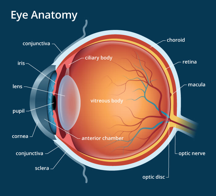
Eye Anatomy: 16 Parts of the Eye & Their Functions - Vision Center The following are parts of the human eyes and their functions: 1. Conjunctiva The conjunctiva is the membrane covering the sclera (white portion of your eye). The conjunctiva also covers the interior of your eyelids. Conjunctivitis, often known as pink eye, occurs when this thin membrane becomes inflamed or swollen.

Eye diagram with labels and functions
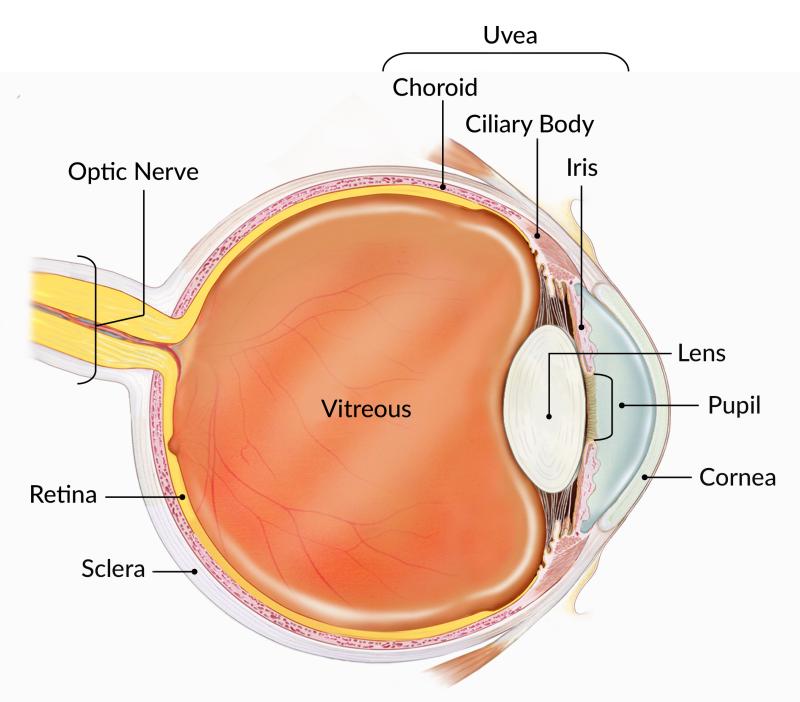
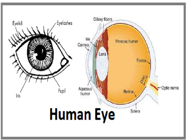
Human Eye: Structure, Diagram, Vision Aspects of Human Eye Human Eye is one of the five sensory organs of the human body. It is the organ that provides us vision, allows us to perceive and differentiate colors, and acts as the biological clock of the human body. Structure of the Human Eye is highly complex. It is made up of a number of nerves and muscles all of which have specific functions. Parts Of The Eye Labeled Diagram Model And Their Function Parts of the eye-labeled diagram model are divided into three groups: the external outer layer, the middle layer, and the inner back layer. The outer layer is responsible for protecting the eye from environmental toxins and debris. The middle layer includes cells that allow light to enter and travel through the back layer to the retina. The Eyes (Human Anatomy): Diagram, Optic Nerve, Iris, Cornea ... - WebMD Just behind the iris and pupil lies the lens, which helps focus light on the back of your eye. Most of the eye is filled with a clear gel called the vitreous. Light projects through your pupil and...
Eye diagram with labels and functions. › ir-receiver-remote-arduinoIR Remote and Receiver with Arduino Tutorial (4 Examples) Aug 23, 2020 · For this example, I connected the output of the IR receiver to digital pin 8 instead of 2. The connections for the character LCD display are shown in the wiring diagram below. Note that you also need a 10 kΩ potentiometer to set the contrast of the display and a 220 Ω resistor to control the brightness of the backlight. › consumers › consumer-updatesConsumer Updates | FDA - U.S. Food and Drug Administration Jul 28, 2022 · The .gov means it’s official. Federal government websites often end in .gov or .mil. Before sharing sensitive information, make sure you're on a federal government site. Eye pattern - Wikipedia In telecommunication, an eye pattern, also known as an eye diagram, is an oscilloscope display in which a digital signal from a receiver is repetitively sampled and applied to the vertical input, while the data rate is used to trigger the horizontal sweep. It is so called because, for several types of coding, the pattern looks like a series of eyes between a pair of rails. Labelling the eye — Science Learning Hub In this interactive, you can label parts of the human eye. Use your mouse or finger to hover over a box to highlight the part to be named. Drag and drop the text labels onto the boxes next to the eye diagram If you want to redo an answer, click on the box and the answer will go back to the top so you can move it to another box.
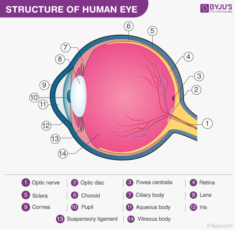
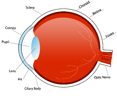
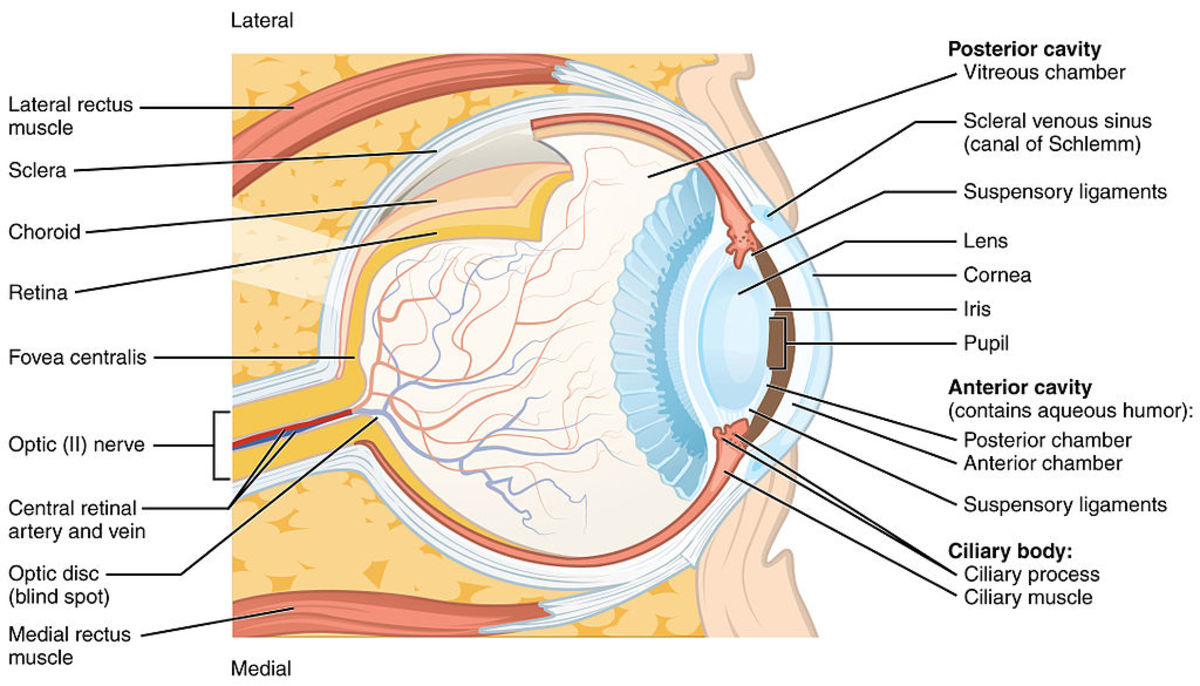
Structure and Function of the Human Eye - ThoughtCo The main parts of the human eye are the cornea, iris, pupil, aqueous humor, lens, vitreous humor, retina, and optic nerve. Light enters the eye by passing through the transparent cornea and aqueous humor. The iris controls the size of the pupil, which is the opening that allows light to enter the lens. Light is focused by the lens and goes ... Eye Diagram With Labels and detailed description - BYJUS A brief description of the eye along with a well-labelled diagram is given below for reference. Well-Labelled Diagram of Eye The anterior chamber of the eye is the space between the cornea and the iris and is filled with a lubricating fluid, aqueous humour. The vascular layer of the eye, known as the choroid contains the connective tissue. Eye Anatomy: Parts of the Eye and How We See Here is a tour of the eye starting from the outside, going in through the front and working to the back. Eye Anatomy: Parts of the Eye Outside the Eyeball The eye sits in a protective bony socket called the orbit. Six extraocular muscles in the orbit are attached to the eye. These muscles move the eye up and down, side to side, and rotate the eye. › about › newsroomEsri Newsroom | Publications, Stories, Articles & Press Coverage Explore thought-provoking stories and articles about location intelligence and geospatial technology. Discover thought leadership content, user publications & news about Esri.
Consumer Updates | FDA - U.S. Food and Drug Administration 28/07/2022 · The site is secure. The https:// ensures that you are connecting to the official website and that any information you provide is encrypted and transmitted securely. en.wikipedia.org › wiki › FluorescenceFluorescence - Wikipedia Fluorescence is the emission of light by a substance that has absorbed light or other electromagnetic radiation.It is a form of luminescence.In most cases, the emitted light has a longer wavelength, and therefore a lower photon energy, than the absorbed radiation. MCAT Eye Anatomy: Eye Structure & Function - Magoosh MCAT Blog Function. Cornea. Outermost lens of the eye responsible for the majority of light refraction. Aqueous humor. Fluid that adds refractive power. Iris/Pupil. Colored tissue that regulates the amount of light entering the eye through the central pupil. *KEY CONCEPT: The pupil dilates in low light conditions and constricts in high light conditions. Cow's Eye Dissection - Eye diagram - Exploratorium The pupil is the dark circle in the center of your iris. It's a hole that lets light into the inner eye. Your pupil is round. A cow's pupil is oval. A tough, clear covering over the iris and the pupil that helps protect the eye. Light bends as it passes through the cornea. This is the first step in making an image on the retina.
Eye: anatomy, physiology and barriers to drug delivery ...
Eye Diagram - an overview | ScienceDirect Topics (a) A perfect square eye diagram, (b) a closed eye diagram due to bandwidth, fiber impairments, noise, and timing jitter. The Q-factor ( q) is also an important system parameter widely used in long-distance optical transmission system design. It is defined as the electrical signal-to-noise ratio before the decision circuit at receiver.

IGCSE Biology 2017: 2.91: Describe the Structure and Function ...
Excel Gauge Chart Template - Free Download - How to Create Move the labels to the appropriate places above the gauge chart. Change the chart title. Bonus Step for the Tenacious: Add a text box with your actual data value. Here is a quick and dirty tip on making the speedometer chart more informative as well as pleasing to the eye. Let’s add a text box that will display the actual value of the pointer.
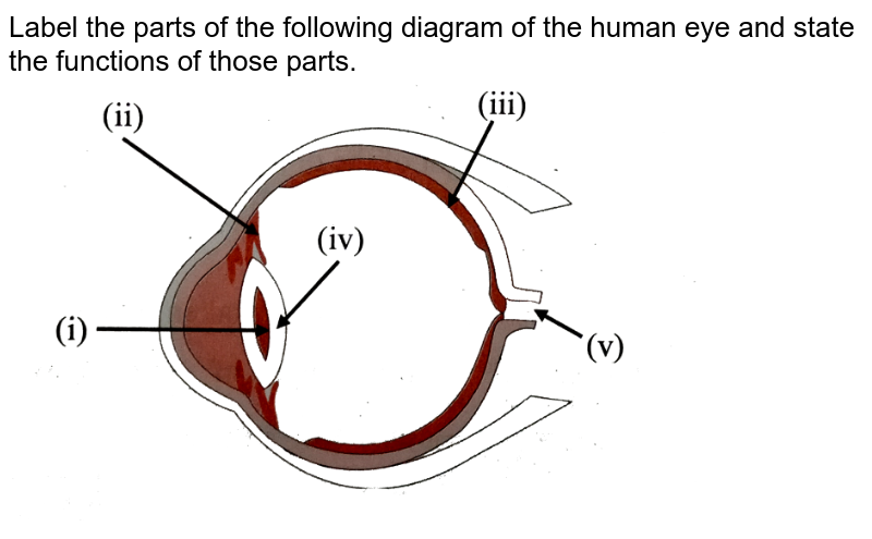
Label the parts of the following diagram of the human eye and ...
The Eye Diagram: What is it and why is it used? The eye diagram is used primarily to look at digital signals for the purpose of recognizing the effects of distortion and finding its source. To demonstrate using a Tektronix MDO3104 oscilloscope, we connect the AFG output on the back panel to an analog input channel on the front panel and press AFG so a sine wave displays. Then we press Acquire.
How the Eyes Work | National Eye Institute
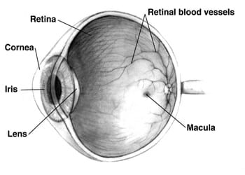
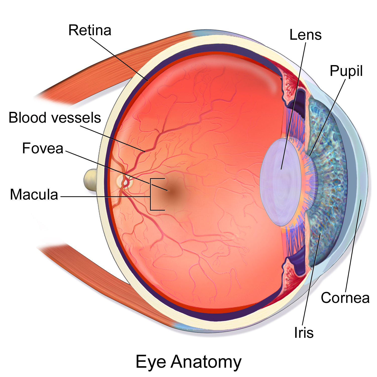
Eye Anatomy Diagram - EnchantedLearning.com Retina - light-sensitive tissue that lines the back of the eye. It contains millions of photoreceptors (rods and cones) that convert light rays into electrical impulses that are relayed to the brain via the optic nerve. Rods - cells the in the retina that sense brightness (they are photoreceptors). Night vision involves mostly rods (not cones).
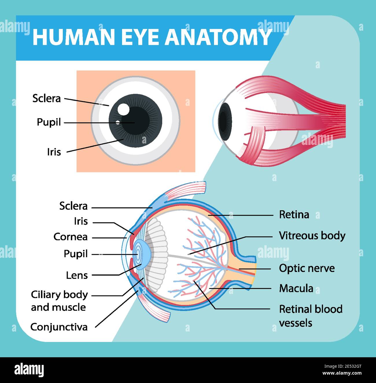
Diagram of human eye anatomy with label illustration Stock ...
Eye anatomy and function - AboutKidsHealth For people with normally functioning eyes, the following sequence takes place: Light reflects off the object we are looking at. Light rays enter the eye through the cornea at the front of the eye. The light passes through a watery fluid (aqueous humor), and enters the pupil to reach the lens.
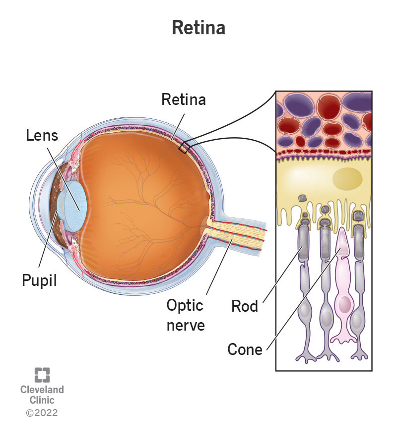
Retina: Anatomy, Function & Common Conditions
Labelled Diagram of Human Eye, Explanation and Function - VEDANTU The basic functions of Rods and Cones are conscious light perception, color differentiation and depth perception. The human eye is capable of distinguishing between about 10 million colors, and it can also detect a single photo. The human eye is a part of the sensory nervous system. Labeled Diagram of Human Eye

Simple eye diagrams | Easy eye diagram | Labeled eye diagram ...
Parts of the Body for Kids: Names & Basic Functions Diagram of Body Parts. External, which means “outside,” describes the body parts that you can see. Take a look at a helpful diagram that labels major external body parts. Download the printable PDF to see it in more detail and print if needed. View & Download PDF
The Human Eye: Anatomy, Structure, Working, Function and Defects
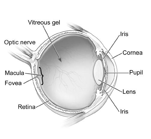
PDF Parts of the Eye - National Institutes of Health To understand eye problems, it helps to know the different parts that make up the eye and the functions of these parts. Here are descriptions of some of the main parts of the eye: ... Handout illustrating parts of the eye Keywords: parts of the eye, eye diagram, vitreous gel, iris, cornea, pupil, lens, optic nerve, macula, retina ...
Structure and Functions of Human Eye with labelled Diagram
The Human Eye - Diagram, Parts, Working, Function and Work of The Lens The human eye operates similar to a digital camera in several ways: Light focuses mainly on the cornea, which acts like a camera lens. The iris controls the light that reaches the eye by adjusting the size of the pupil, and thus it functions like the diaphragm of a camera. The lens of the eye is located behind the pupil, and it focuses light.
Eye Anatomy: Parts of the Eye and How We See - American ...
dusk.geo.orst.edu › gis › lec11_12GEO 465/565 - Lectures 11 and 12 - Spatial Analysis Before the more detailed analysis, it is useful to create a diagram or flow chart of the layers and analysis functions you will use. For this analysis you will follow the steps shown in the flow chart below. First, you will query the Land use layer to create a new layer that contains only residential areas.

Human eye Diagram | Quizlet
Human Eye Diagram, How The Eye Work -15 Amazing Facts of Eye First, light rays enter the eye through the cornea, the clear front "window" of the eye. The dome shaped cornea bends light to help the eye focus. From the cornea, the light passes through an opening called the pupil. The amount of light passing through is controlled by the iris, or the colored part of your eye.

Anatomy of the eye - Moorfields Eye Hospital
Label the microscope — Science Learning Hub 08/06/2018 · All microscopes share features in common. In this interactive, you can label the different parts of a microscope. Use this with the Microscope parts activity to help students identify and label the main parts of a microscope and then describe their functions.. Drag and drop the text labels onto the microscope diagram. If you want to redo an answer, click on the box and …
describe the anatomy of the eye and the function of each part pages 310 14
Parallel categories diagram in Python - Plotly Multi-Color Parallel Categories Diagram¶. The color of the ribbons can be specified with the line.color property. Similar to other trace types, this property may be set to an array of numbers, which are then mapped to colors according to the the colorscale specified in the line.colorscale property.. Here is an example of visualizing the survival rate of passengers in the titanic …

draw a neat labelled diagram of the human eye and mention the ...
Labelling the eye — Science Learning Hub In this activity, students use online or paper resources to identity and label the main parts of the human eye. By the end of this activity, students should be able to: identify the main parts of the human eye; describe the functions of the different parts of the human eye. Download the Word file (see link below).
The Human Eye: A Diagram - FamilyConnect
alex.state.al.us › plansALEX | Alabama Learning Exchange Throughout this lesson, students will discover how the lens in your eye helps focus light. First, students will discuss the parts of the eye and how these parts work together to allow us to see. Then, students will use a clear plastic bag filled with water to create a model of an eyeball to investigate how the lens in your eye helps focus light.

Simple eye diagrams | Easy eye diagram | Labeled eye diagram ...
Human Heart - Anatomy, Functions and Facts about Heart - BYJUS The human heart functions throughout a person’s lifespan and is one of the most robust and hardest working muscles in the human body. Besides humans, most other animals also possess a heart that pumps blood throughout their bodies. Even invertebrates such as grasshoppers possess a heart like pumping organ, though they do not function the same way a human heart …
Parts of the Eye & Their Function | Robertson Optical and ...
Human Eye Ball Anatomy & Physiology Diagram - eMedicineHealth A ring of muscular tissue, called the ciliary body, surrounds the lens and is connected to the lens by fine fibers, called zonules. Together, the lens and the ciliary body help control fine focusing of light as it passes through the eye. The lens, together with the cornea, functions to focus light onto the retina. Manage Diabetes in 10 Minutes

Labeled Eye Diagram | Science Trends
Control Unit Installation and Operation Guide Please Read between any Eye QS control unit and any other power supply, including another GRAFIK Eye QS control unit. Refer to the QS Link Power Draw Units specification submittal (Lutron P/N 369405) for more information concerning PDUs. 1234 12 ABC 123456LN Example: Emergency lighting interface (maximum 1) Note: The GRAFIK Eye QS control unit

Label the Eye Diagram | Quizlet
Eye Anatomy: A Closer Look At the Parts of the Eye - All About Vision The iris of the eye functions like the diaphragm of a camera, controlling the amount of light reaching the back of the eye by automatically adjusting the size of the pupil (aperture). The eye's crystalline lens is located directly behind the pupil and further focuses light.
![Cross sectional diagram of human eye [1]. | Download ...](https://www.researchgate.net/publication/276541864/figure/fig1/AS:612895498964992@1523137082339/Cross-sectional-diagram-of-human-eye-1.png)
Cross sectional diagram of human eye [1]. | Download ...
Generate eye diagram - MATLAB eyediagram - MathWorks eyediagram(x,n) generates an eye diagram for signal x, plotting n samples in each trace.The labels on the horizontal axis of the diagram range between –1/2 and 1/2. The function assumes that the first value of the signal and every nth value thereafter, occur at integer times.

Parts and Functions of the Human Eye Diagram | Quizlet
Eye Muscles - All About Vision The main function of the extraocular eye muscles is to control eye movement and eye alignment. They are different from the intrinsic eye muscles, which enable the eye to focus on near objects and control how much light enters the eye. Extraocular eye muscles and their functions

Functions and Anatomy of the Eye | Health and physical ...
eye labeling Diagram | Quizlet sclera. Tough white out covering of the eyeball. choroid. Middle layer of the eye (between the retina and the sclera) that contains the blood vessels that nourish the eye and cornea. iris. colored layer that dilates and constricts to allow in more or less light. ciliary body. structure on each side of the lens that connects the choroid and iris.
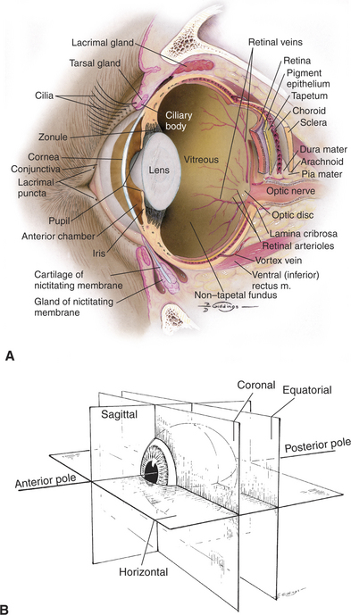
Structure and Function of the Eye | Veterian Key
Diagram of the Eye - Lions Eye Institute To understand the eye and its functions, it's important to understand how the eye works, see below diagrams for both the external eye and the internal eye. The External Eye Instructions Click the parts of the eye to see a description for each. Hover the diagram to zoom. The Internal Eye Instructions

Anatomy of the Eye | Human eye diagram, Eye anatomy, Eye ...
Parts of Stereo Microscope (Dissecting microscope) – labeled diagram ... If you would like to learn optical components of a compound microscope, please visit Compound Microscope Parts – Labeled Diagram and their Functions, and this article. How to use a stereo (dissecting) microscope. Follow these steps to put your stereo microscopes in work: 1. Set your microscope on a tabletop or other flat sturdy surface where ...

Module 1: Labeled Diagram of the Eye | Diagram of the eye ...
Anatomy of the eye: Quizzes and diagrams | Kenhub Take a look at the diagram of the eyeball above. Here you can see all of the main structures in this area. Spend some time reviewing the name and location of each one, then try to label the eye yourself - without peeking! - using the eye diagram (blank) below. Unlabeled diagram of the eye. Click below to download our free unlabeled diagram of ...

Draw a neat labeled diagram of human eye and explain the ...
PDF Eye Anatomy Handout - National Institutes of Health of light entering the eye. Lens: The lens is a clear part of the eye behind the iris that helps to focus light, or an image, on the retina. Macula: The macula is the small, sensitive area of the retina that gives central vision. It is located in the center of the retina. Optic nerve: The optic nerve is the largest sensory nerve of the eye.
Eye Anatomy: A Closer Look At the Parts of the Eye
Esri Newsroom | Publications, Stories, Articles & Press Coverage Explore thought-provoking stories and articles about location intelligence and geospatial technology. Discover thought leadership content, user publications & news about Esri.

Structure And Functions of the different part of the Human ...
Structure and Functions of Human Eye with labelled Diagram - BYJUS Structure and Functions of Human Eye with labelled Diagram Biology Biology Article Structure Of Eye Structure of the Eye The eye is one of the sensory organs of the body. In this article, we shall explore the anatomy of the eye The structure of the eye is an important topic to understand as it one of the important sensory organs in the human body.

Human Eye Anatomy - Parts of the Eye Explained | Eye anatomy ...
The Eyes (Human Anatomy): Diagram, Optic Nerve, Iris, Cornea ... - WebMD Just behind the iris and pupil lies the lens, which helps focus light on the back of your eye. Most of the eye is filled with a clear gel called the vitreous. Light projects through your pupil and...

Draw a labelled diagram of V.S. of the human eye and write ...
Parts Of The Eye Labeled Diagram Model And Their Function Parts of the eye-labeled diagram model are divided into three groups: the external outer layer, the middle layer, and the inner back layer. The outer layer is responsible for protecting the eye from environmental toxins and debris. The middle layer includes cells that allow light to enter and travel through the back layer to the retina.
Eye Anatomy Diagram - EnchantedLearning.com
Human Eye: Structure, Diagram, Vision Aspects of Human Eye Human Eye is one of the five sensory organs of the human body. It is the organ that provides us vision, allows us to perceive and differentiate colors, and acts as the biological clock of the human body. Structure of the Human Eye is highly complex. It is made up of a number of nerves and muscles all of which have specific functions.

15.5 Vision – Anatomy & Physiology
Anatomy and Structure of the Human Eye (With Diagrams ...
Eyes (Anatomy): Overview, Parts and Functions | Biology ...

function parts of the eye Diagram | Quizlet
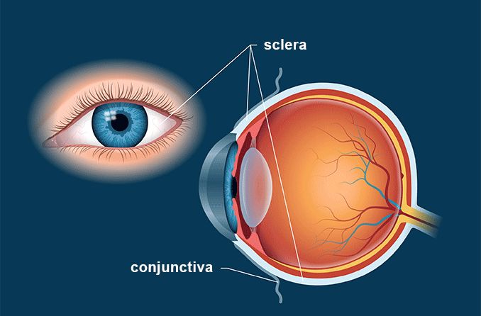
Sclera | White of the Eye - Definition and Detailed Illustration

Eye Anatomy and How the Eye Works
Macula of retina - Wikipedia

Structure and Functions of Human Eye with labelled Diagram
Post a Comment for "39 eye diagram with labels and functions"